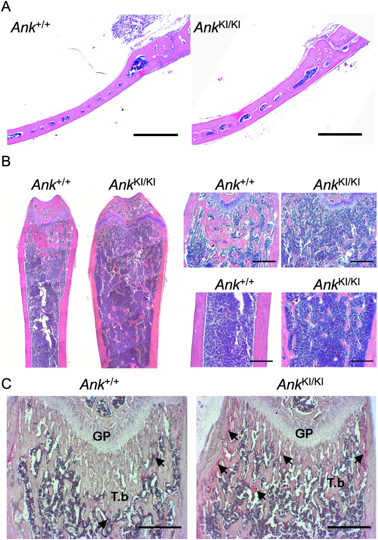
FIG. 3.
Histology of Ank +/+ and Ank KI/KI male mice. (A) Calvariae from 10-wk-old mice (H&E). (B) H&E staining of 10-wk-old mice: femurs (left panel), metaphyses (top right), and diaphyses (bottom right). (C) TRACP staining of femurs from 4-wk-old mice. Arrows indicate TRACP+ cells. GP, growth plate; T.b, trabecular bone. Scale bar = 500 μm.

