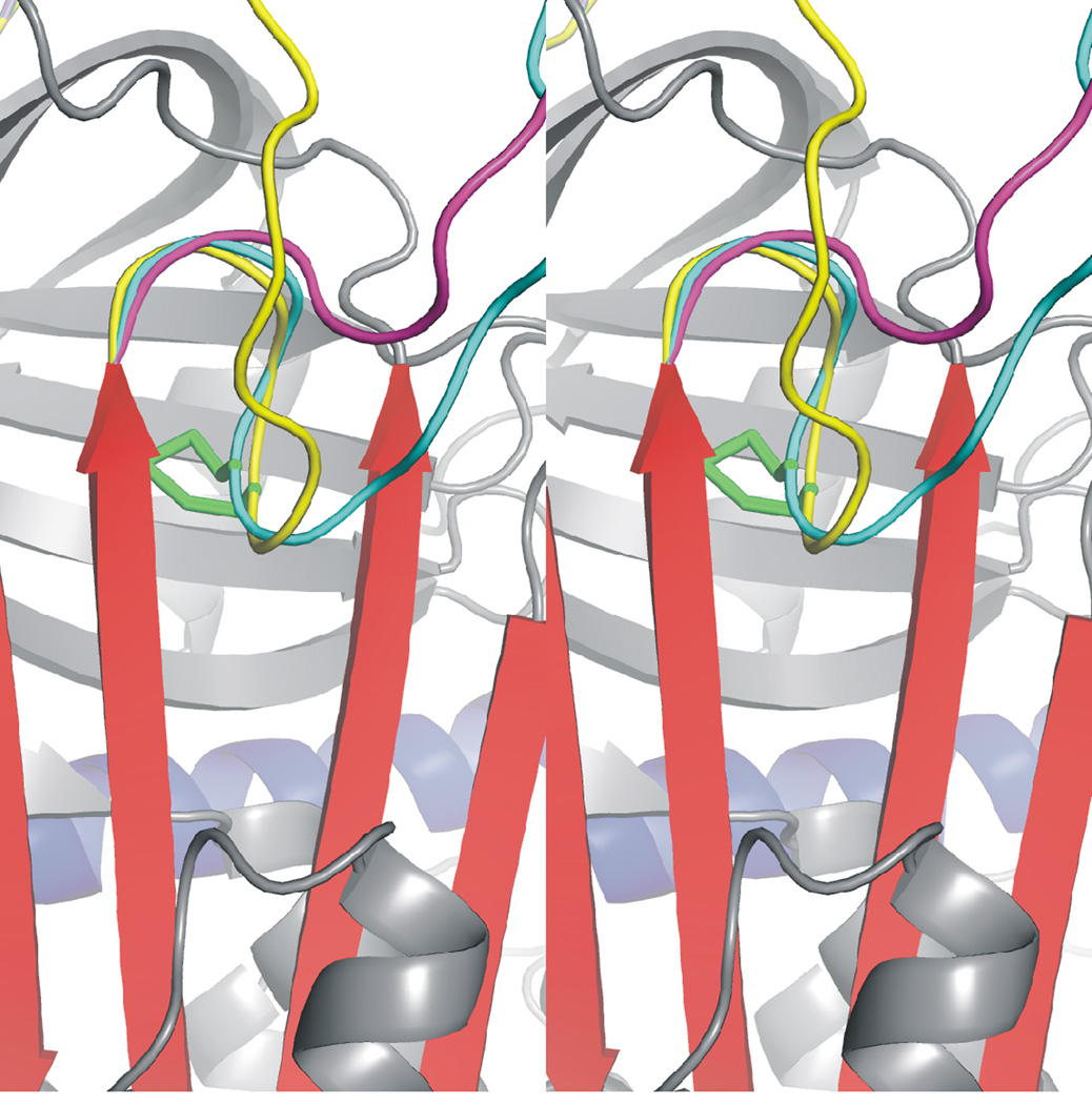
Figure 2.
Internal disulphide bond prevents the expulsion of the hinge from β-sheet A. Close up of the internally oriented disulphide bond (AT coloured and oriented as in Fig. 1), between the hinge region and strand 5A. The RCL before energy minimization is coloured in yellow, and the stereo view allows the depth of the disulphide bond (green) to be appreciated. Energy minimization of the RCL results in the full expulsion of the hinge in the absence of the disulphide bond (magenta), while no expulsion is permitted for the variant after the same energy minimization regime (cyan).

