Figure 4.

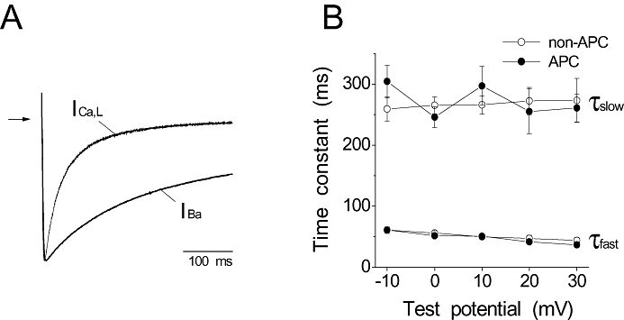
Effects of in vivo APC on the voltage-dependent inactivation of the cardiac L-type Ca channel. (A) Representative traces of ICa,L and IBa recorded at 0 mV from a holding potential of −80 mV using Ca2+ and Ba2+ as charge carriers, respectively, are shown. The currents were scaled and superimposed. Arrow indicates zero current level. Ba2+ as the charge carrier removed Ca2+-dependent inactivation of the L-type Ca channel. (B) The fast and slow time constants of the inactivation kinetics of IBa in the non-APC and APC groups are plotted against test potentials. No significant differences were observed in τfast and τslow between the non-APC and APC groups (n = 10 per group).
