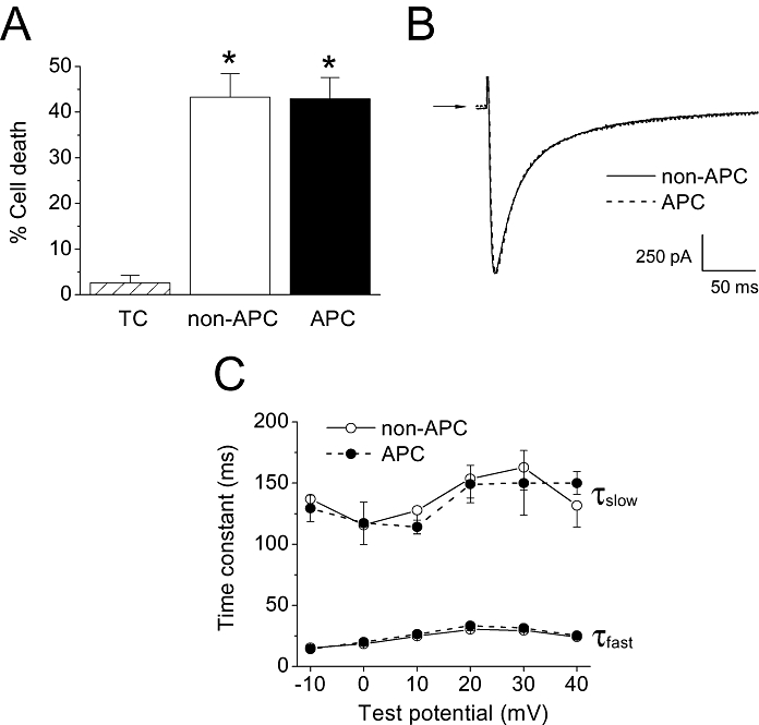
Figure 9.

Effects of in vivo APC on the Dahl S strain of rats. (A) Cell survival study. Oxidative stress significantly increased the percentage of cell death in the non-APC group compared with the time control (TC) group. In the APC group, cell survival following oxidative stress was not significantly different from the APC group, indicating resistance to cardioprotection in the Dahl S rats. *P < 0.05 vs. TC. n = 5–9 per group. (B) Representative whole-cell ICa,L traces from Dahl S myocytes in the non-APC and APC groups. Currents were recorded at a test potential of 0 mV from a holding potential of −80 mV. The traces shown were superimposed. Arrow indicates zero current level. (C) ICa,L inactivation kinetics. The fast and slow time constants of current inactivation in myocytes from the non-APC and APC groups showed no significant differences in the Dahl S rats (n = 9 per group).
