
Figure 2.
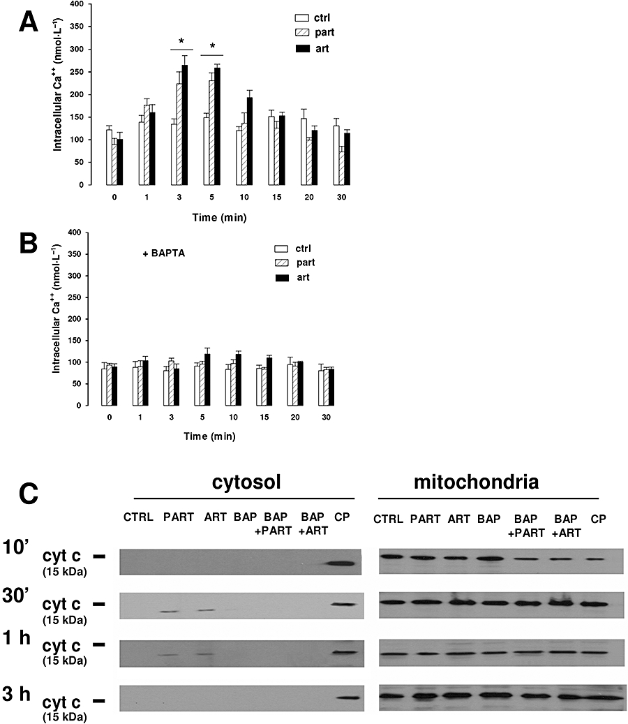
Effects of parthenolide and artemisinin on [Ca++]i. (A) Cells were grown on sterile glass coverslips for 24 h, washed with PBS and incubated for 10 min in HEPES-Ca buffer containing 10 µmol·L−1 FURA-AM, in the absence (ctrl) or presence of either parthenolide (part, 10 µmol·L−1) or artemisinin (art, 10 µmol·L−1). Fluorescence was detected throughout the first 30 min, as described under Methods section. Measurements were performed in duplicate and data are presented as means ± SE (n = 6). Significance of each drug versus ctrl: *P < 0.005. (B) Cells were grown on sterile glass coverslips as described previously, incubated for 1 h with 10 µmol·L−1 BAPTA-AM, then washed with PBS, and subjected to the same experimental procedure described in panel A. Measurements were performed in duplicate and data are presented as means ± SE (n = 4). (C) Cells were incubated for different times in the absence (CTRL) or in the presence of either parthenolide (PART, 10 µmol·L−1) or artemisinin (ART, 10 µmol·L−1). When indicated, cells were pre-loaded with the Ca++ chelator BAPTA (BAP) (see above). As a positive control, several samples were previously incubated for 24 h with the pro-apoptotic drug camptothecin (CP, 10 nmol·L−1). Cytosolic and mitochondrial fractions were separated and subjected to Western blotting analysis for cytochrome c, as described under Methods section. The Figure is representative of three experiments with similar results. BAPTA-AM, 1,2-bis(2-aminophenoxy)ethane-N,N,N′,N′-tetraacetic acid-acetoxymethylester; PBS, phosphate buffer saline.