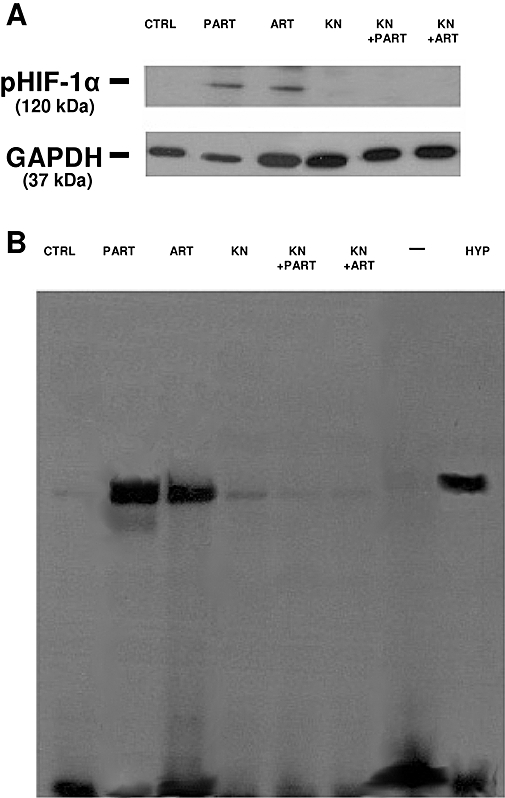
Figure 7.

Effect of parthenolide, artemisinin and KN93 on HIF-1α phosphorylation and nuclear translocation. HT29 cells were incubated for 3 h in the absence (CTRL) or in the presence of either parthenolide (PART, 10 µmol·L−1) or artemisinin (ART, 10 µmol·L−1), alone or together with KN93 (KN, 10 µmol·L−1), then the following investigations were performed. (A) Western blotting detection of phospho(Ser)-HIF-1α (pHIF-1α). Whole cell lysates were incubated with an anti-HIF-1α antibody. Subsequently the immunoprecipitated proteins were separated by SDS-PAGE and probed with an anti-phospho(Ser) antibody as described in the Methods section. The expression of GAPDH was used as a control of equal protein loading. The Figure is representative of three experiments with similar results. (B) EMSA detection of HIF-1α nuclear translocation. EMSA was performed on nuclear extracts as detailed in Methods section. In each experiment, one lane was loaded with double-distilled water (–) instead of cellular extracts. The lane marked with HYP was loaded with nuclear extracts obtained from HT29 cells incubated for 3 h in a humidified hypoxic atmosphere (3% O2, 5% CO2, 37°C), to achieve a maximal HIF-1α activation. The Figure is representative of three experiments with similar results. EMSA, electrophoretic mobility shift assay; GAPDH, glyceraldehyde-3-phosphate dehydrogenase; HIF-1α, hypoxia-inducible factor-1α.
