
Figure 4.
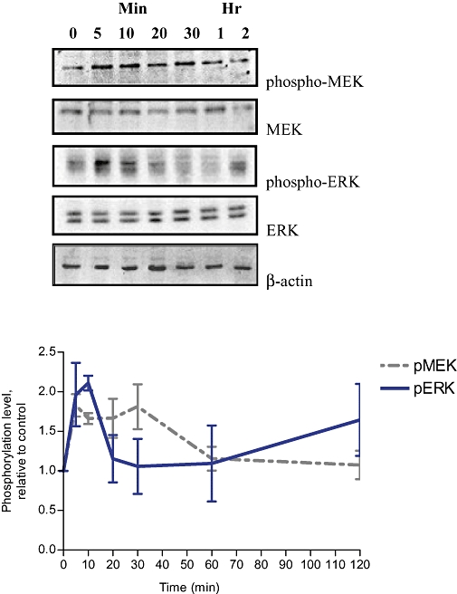
Time course of MEK and ERK activation by Rg1 in MCF-7 cells. MCF-7 cells were stimulated with 1 pmol·L−1 Rg1 for various lengths of time (0, 5 min, 10 min, 20 min, 30 min, 1 h and 2 h). After SDS-PAGE, blots were immunoblotted with specific antibodies. The signals were detected and quantified by the Lumi-Imager. Upper panel shows the immunoblot against phospho-MEK, MEK, phospho-ERK, ERK and β-actin. Lower panel shows the degree of phosphorylation which is calculated first as a ratio of pMEK to MEK or a ratio of pERK to ERK and finally expressed as a ratio to the basal reading where time 0 (untreated as basal) equals 1. The results shown were obtained from three independent experiments and expressed as mean ± SEM; n = 3.