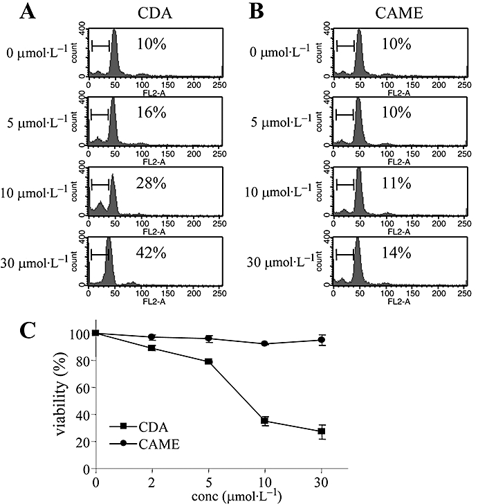
Figure 3.

Effects of CDA and CAME on cell apoptosis and cell viability of primary Th cells. Single suspensions of CD4+ Th cells were stimulated with anti-CD3 (1 µg·mL−1) and anti-CD28 (1 µg·mL−1) for 24 h in the presence of different concentrations of CDA (A) or CAME (B); numbers in the records show the percentage of the cells in sub-G1 (using ModFit software). (C) Cells treated with either CDA or CAME for 24 h were incubated with MTT solution, and colorimetric changes were determined by elisa reader. Data are given as means ± SD, n= 3. CAME, p-coumaryl alcohol-γ-O-methyl ether; CDA, p-coumaryl diacetate; conc, concentration; elisa, enzyme-linked immunosorbent assay; MTT, dimethylthiazol diphenyltetrazolium bromide; Th cell, T helper cell.
