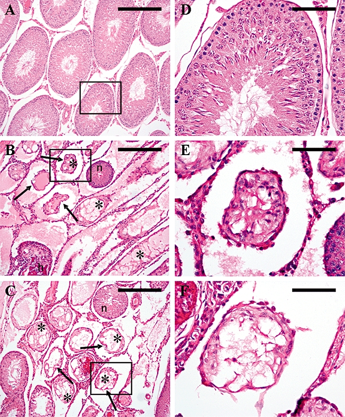
Figure 9.

Seminiferous tubule histology at 2 months following a single s.c. injection of kisspeptin-54 (50 nmol) or GnRH (50 nmol) in the adult male rat. Cross sections of testicular tubules assessed by light microscopy on H&E-stained sections. (A) Representative section from a control animal showing tubules with normal maturation to late-stage elongated spermatids and a few spermatozoa. (B) Representative section of an animal treated with a single s.c. injection of 50 nmol kisspeptin-54 showing tubules with loss of seminiferous tubular structure (arrows) with vacuolation and atrophy (asterisk) and tubular hyalinization (h) as common features. The section also shows an apparently normal seminiferous tubule alongside the degenerated tubules (n). (C) Representative section of an animal treated with a single s.c. injection of 50 nmol GnRH showing the same features as described for kisspeptin-54 in (B). (Original magnifications ×100. Scale bar represents 100 µm). (D–F) Higher magnification of (A–C), to show a single tubule (boxed section) from each treatment group. (Original magnifications ×400. Scale bar represents 25 µm). GnRH, gonadotrophin-releasing hormone; H&E, haematoxylin and eosin.
