
Figure 3.
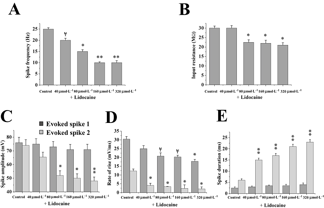
Effect of lidocaine upon evoked spike parameters. Histograms showing the effects of lidocaine (40–320 µmol·L−1; 15 min) on neuronal membrane properties and evoked spike parameters: (A) mean spike firing frequency (n= 5), (B) input resistance (n= 7), (C) first and second spike amplitude (n= 5), (D) first and second spike rate of rise (n= 5) and (E) first and second spike duration (n= 5). Data in A, C–E were all calculated following a +1.5 nA, 160 ms current pulse; evoked first spike and evoked second spike. All responses were elicited from resting potential (∼−83 mV). Note that the effects of carisbamate (see Figs 1 and 2) were comparable to those of lidocaine, on spike frequency, second spike amplitude, rate of rise and duration, suggesting a common mechanism of action; also, input resistance was significantly reduced by lidocaine at concentrations ≥80 µmol·L−1. ¥ indicates a trend assessed by P < 0.1; * and ** indicate differences from control at P < 0.05 and P < 0.01 respectively. Significance accepted at P < 0.05. Data shown are means ± SEM for all histograms.