Figure 8.

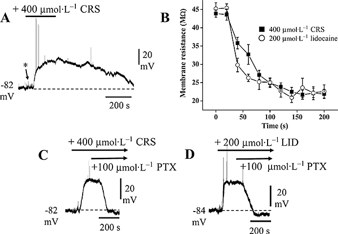
Effects of carisbamate (CRS) and lidocaine (LID) in Cl−-loaded cells. Chart records showing changes in membrane potential and input resistance produced by CRS and LID in cells recorded at normal resting potential, by using 2 mol·L−1 KCl-filled microelectrodes [* indicates increase in baseline ‘noise’ (cf. Figure 1B) probably due to spontaneous, depolarizing GABA release]. (A) A brief (150 s) application of CRS (400 µmol·L−1) produced a depolarization and slow recovery on washout; note the spontaneous firing induced at the onset of the depolarizing response (action potentials truncated by chart recorder). (B) Comparison of the input resistance changes produced by 400 µmol·L−1 CRS and 200 µmol·L−1 LID recorded in Cl−-loaded cells (as in a,c,d) (data points are means ± SEM, n= 4 for both CRS and LID). (C) CRS response recorded in a different neurone: application of picrotoxin (PTX; 100 µmol·L−1) in the continued presence of CRS reversed the CRS-induced depolarization. (D) Application of LID (200 µmol·L−1) in a different cell evoked a similar depolarization that was also reversed by applying PTX (in the presence of LID).
