
Figure 3.
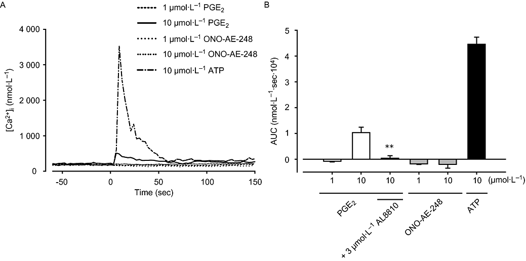
Effect of prostaglandin E2 (PGE2) and ONO-AE-248 on [Ca2+]i. (A) Liver myofibroblasts cultured on glass coverslips were stimulated with PGE2 and ONO-AE-248 (1 or 10 µmol·L−1), or ATP (10 µmol·L−1). Changes in [Ca2+]i were measured using Ca2+ fluorescent dye fura-2. (B) The area under [Ca2+]i-time curve after stimulation (AUC: 0–2 min). Data are presented as mean ± SEM of 52–110 cells from four separate experiments. **P < 0.01 compared with 10 µmol·L−1 PGE2-treated cells. AUC, area under curve.