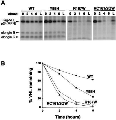
Figure 2.
Pulse/chase analysis of pVHL. (A) 786-O cells stably expressing either WT or point mutant (containing amino acids substitutions indicated above each panel) Flag-tagged pVHL were pulsed with [35S]methionine for 2 h. Cells were then chased with unlabeled media for 0, 2, 4, or 6 h, or for 6 h with lactacystin (L), as indicated above each autoradiograph panel. Flag-VHL proteins were immunoprecipitated with anti-Flag agarose beads (Sigma) and complexes were separated by SDS-PAGE and visualized by fluorography. Positions of Flag-VHLp24(MPR), elongin B, and elongin C proteins are indicated by arrowheads to the left. (B) Radioactive bands in A were quantified and graphically represented.

