Figure 3.

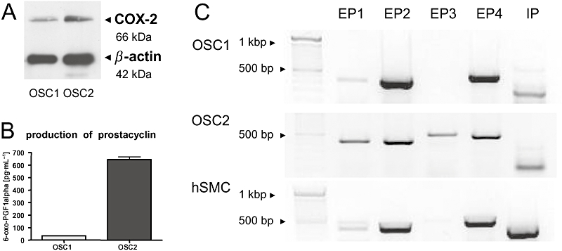
Characterization of the COX2/PG system in OSC1 and OSC2 cells. (A) As shown by immunoblotting, COX2 was more abundantly expressed in OSC2 cells than in OSC1 cells. One representative immunoblot out of three is shown. (B) To determine the amount of PGs secreted by OSC1 and OSC2 cells, the prostacyclin metabolite, 6-oxo-PGF1α, indicative of PG synthesis was determined in the supernatant. OSC2 cells produced approximately 12-fold more prostacyclin than OSC1 cells (n = 3, mean ± SEM). (C) Expression of PG receptors by OSC1 and OSC2 cells was assessed by RT-PCR. Human smooth muscle cells were examined as control. All cells expressed EP1, EP2, EP4 and IP receptors. Only OSC2 cells additionally expressed marked amounts of EP3 receptor. Receptor bands were found at the expected sizes, EP1/2 (450 bp), EP3/4 (580 bp), IP (380/520 bp). Shown are the results of one representative experiment out of three. COX2, cyclooxygenase-2; hSMC, human vascular smooth muscle cell; OSC, oesophageal squamous cell; PG, prostaglandin; RT-PCR, reverse transcriptase polymerase chain reaction.
