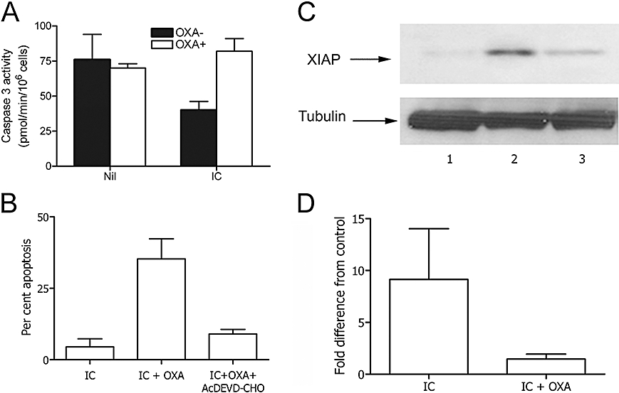
Figure 2.

Oxaprozin (OXA)-induced monocyte apoptosis was dependent on caspase-3 activity and XIAP expression. (A) Human monocytes cultured in the absence (Nil) or presence (IC) of IC were exposed to medium (solid columns), or OXA (open columns). Then, caspase-3 activity was determined spectrophotometrically on whole-cell lysates. Results are expressed as the mean ± 1 s.d., n = 4. Without IC: OXA− versus OXA+: ns. With IC: OXA− versus OXA+: P < 0.01. (B) Microscopic analysis of the apoptosis of human monocytes after acridine orange staining. Monocytes were cultured with IC alone (IC), IC plus OXA (IC + OXA) and IC plus OXA in presence of the caspase-3 inhibitor Ac-DEVD-CHO (IC + OXA + Ac-DEVD-CHO). Data are expressed as mean ± 1 s.d., n = 3. The % spontaneous apoptosis in the absence or presence of OXA (50 µmol·L−1): 38 ± 6 and 36 ± 6, mean ± 1 s.d., n = 3. IC versus. IC + OXA: P < 0.01, IC + OXA versus IC + OXA + Ac-DEVD-CHO: P < 0.01. (C) Western blot: monocyte lysates after incubation of the cells in medium (lane 1), IC (lane 2), IC plus OXA (lane 3). (D) Densitometric analysis of Western blot. Data are expressed as the mean ± 1 s.d., n = 3, and presented as fold difference from control (medium). Fold difference from control of OXA alone: 0.17 ± 0.65. Medium versus IC: P < 0.05; IC versus IC + OXA: P < 0.05. IC, immune complex; XIAP, X-linked mammalian inhibitor of apoptosis protein.
