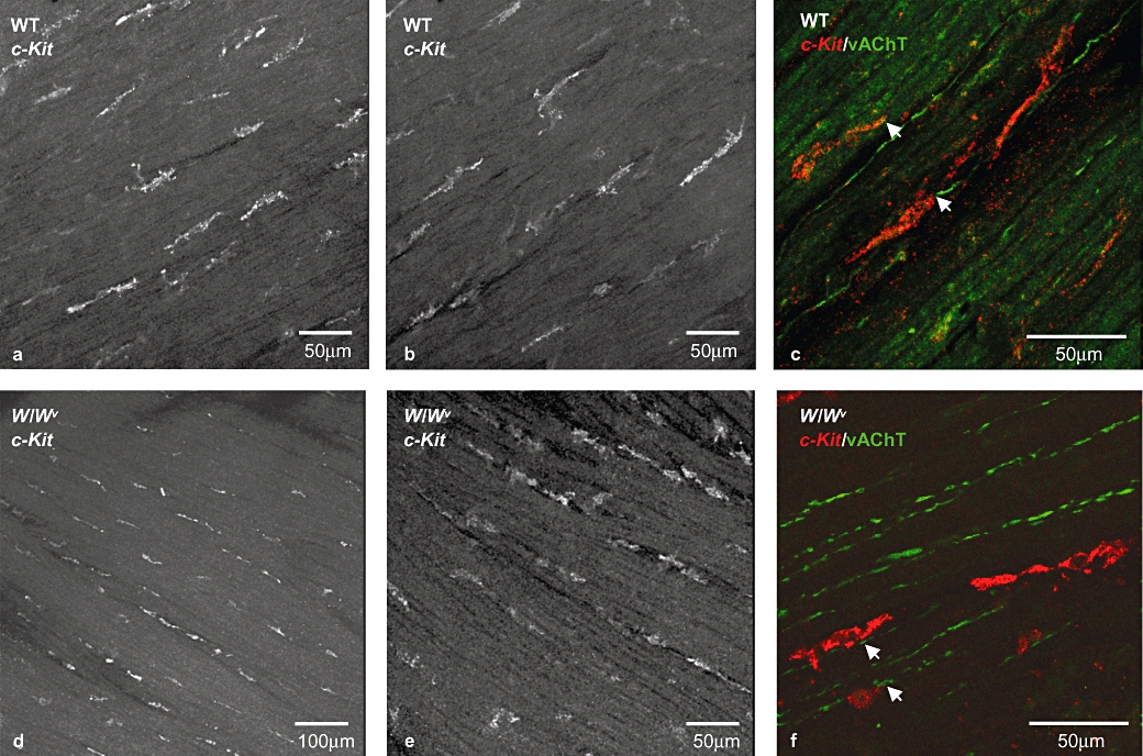
Figure 1.

c-Kit-positive cells in wild-type and W/Wv bladder. c-Kit-positive cells in whole-mount preparations of wild-type (a,b) and W/Wv (d,e) mouse detrusor. Immunoreactive cells were elongated with several small lateral branches and were orientated parallel to the detrusor muscle fibres. Double-labelling with an antibody to vesicular acetylcholine transferase (vAChT, green) and anti-c-Kit (red) revealed close proximity between interstitial cells of Cajal and cholinergic neurons in both tissue types (c,f). All images are projections of a stack of optical sections captured with a confocal microscope.
