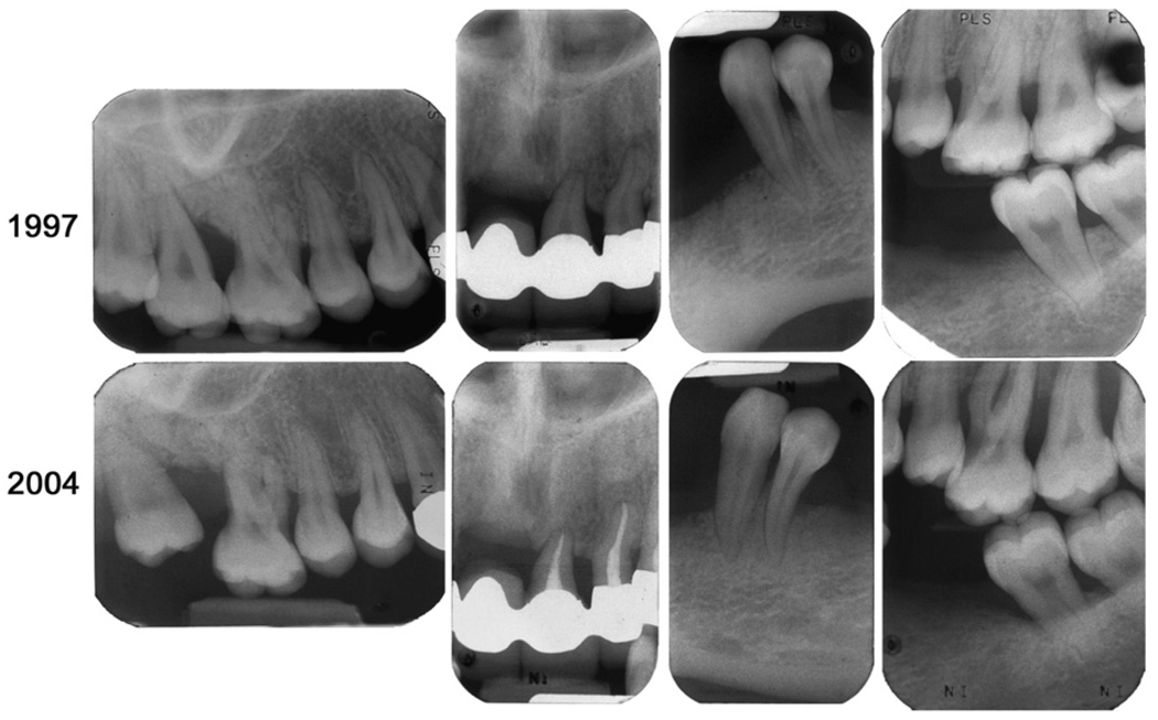
Figure 2.
Selective radiographic comparison of the bone levels 1 year after her initial treatment was completed (1997) and at the most recent complete survey (2004). Note the progression of bone loss around all of the maxillary molars, whereas some teeth with minimal bone support (remaining maxillary incisors) show no major alterations.

