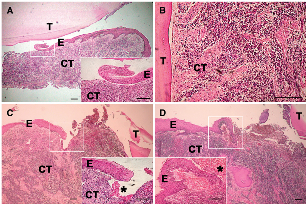
Figure 5.
Histologic assessment of periodontal tissue of maxillary molars of the patient with KS. The teeth were removed gently with forceps, with a small amount of gingival tissue attached, and processed for routine histology. Atypical junctional/pocket epithelium was present in all specimens that showed proper morphology (three separate blocks shown in A, C, and D). In two specimens (A and C), the epithelium showed a blunt end with no attachment to the tooth surface. In one specimen (D), the end of the epithelium resembled epithelium migrating into a wound. Red blood cells (*) were visible in many samples, indicating trauma or spontaneous bleeding (as complained about by the patient). Heavy chronic inflammation was present in all specimens examined (A through D). The inflammatory infiltrates were often approaching the root surface deep in the connective tissue without the epithelial enclosure that is seen in classic periodontitis specimens (B, which represents a deeper area of the same specimen shown in A). Considering the extent of inflammation, the rete ridges were poorly developed or non-existent with signs of epithelial separation from the connective tissue (A, C, and D). T = tooth; E = epithelium; CT = connective tissue. Bars = 200 µm.

