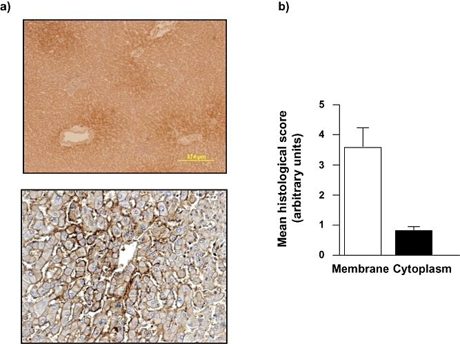
Figure 2.

Normal liver in the absence of hepatocellular carcinoma (HCC) is characterized by zonal aquaporin (AQP) 9 distribution and is predominantly localized to the plasma membrane. (a) Representative immunohistochemical (IHC) images of normal liver (NL) section following IHC staining using an anti-human AQP 9 antibody. Note the zonal distribution (upper panel) and the membrane localization (lower panel). (b) Cumulative scoring analysis of membrane vs. cytoplasmic staining for AQP 9 in hepatocytes in NL. Values are means ± standard error of the mean of five separate fields scored independently by two different investigators; n = 2 separate samples. *P < 0.05
