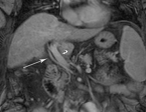
Figure 1.

Coronal enhanced T1-weighted magnetic resonance image shows aberrant right hepatic arterial anatomy. As the replaced right hepatic artery (straight arrow) courses to the liver hilum, it is seen to be caudal and lateral to the portal vein (curved arrow), reflecting a variant origin from the superior mesenteric artery
