Figure 7.

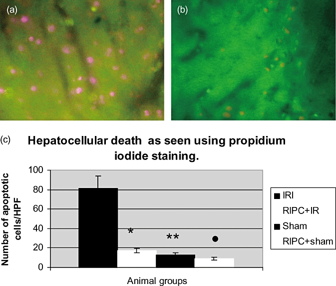
(a) Hepatocellular cell death in ischemia reperfusion injury (IRI) using propidium iodide staining (IVM). The dead cells appear pink stained by propidium iodide. The number of cells divided by the surface area of the field above gives the number of cells/mm2. (b) Hepatocellular cell death seen in remote ischemic preconditioning (RIPC)+IR using IVM. (c) Hepatocellular cell death in the preconditioned group (RIPC+IRI) was significantly less compared with the non-preconditioned (IRI) group. Values expressed as mean ± SEM. *P < 0.05 (IR/RIPC+IR), **P < 0.05 (IR/Sham), •, P < 0.05 (IR/RIPC+Sham)
