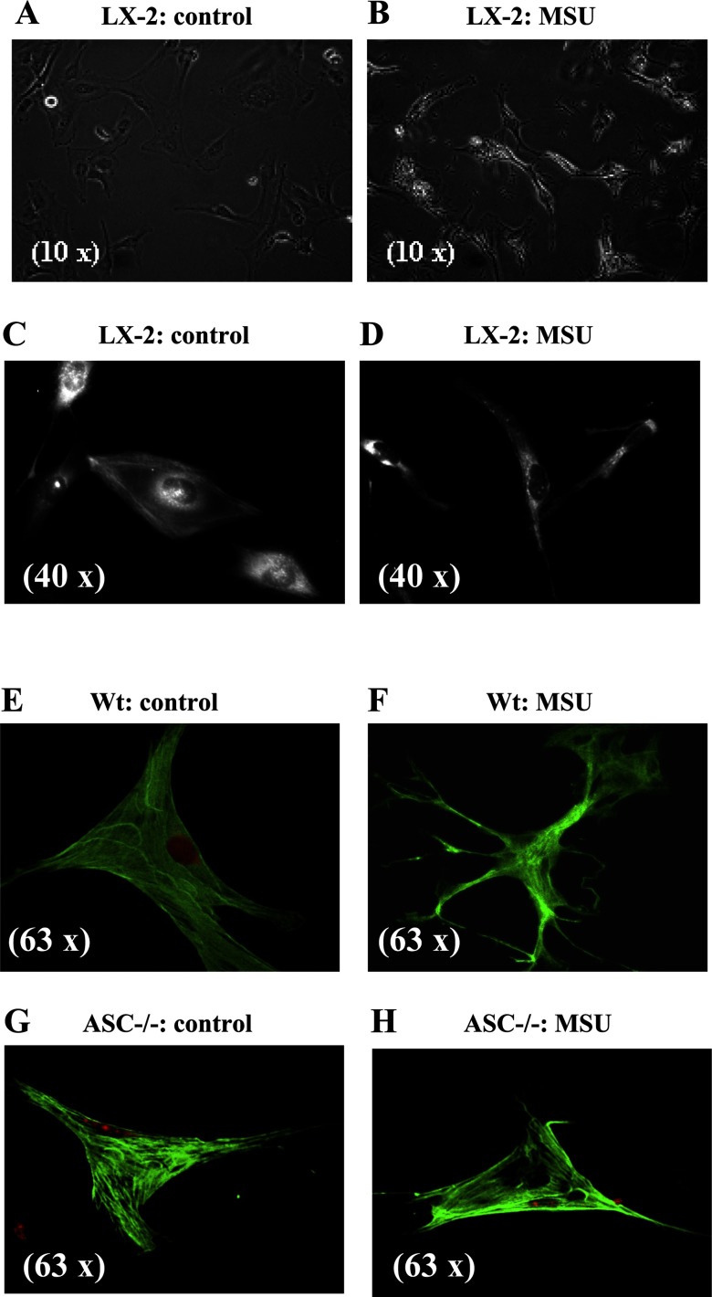
Fig. 2.
MSU (100 μg/ml) induces actin reorganization and stellation of LX-2 cells and primary mice HSC from wild-type but not ASC−/− mice. A: phase-contrast images of LX-2 cells in culture have a flat cuboidal shape. B: phase-contrast images of LX-2 cells with MSU that induces actin reorganization and stellation. C: α-smooth muscle actin (SMA) staining of LX-2 cells shows a flat cuboidal shape. D: after exposure to MSU, this becomes more elongated. E and F: images of primary HSC from wild-type mice with α-SMA staining show actin reorganization after exposure to MSU. G and H: images of primary HSC from ASC−/− mice with α-SMA staining show absence of actin reorganization after exposure to MSU.

