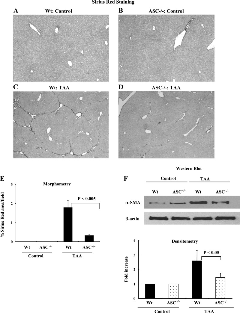
Fig. 6.
Mice lacking ASC (ASC−/−) have reduced collagen deposition in a thioacetamide (TAA) model of liver fibrosis. A and B: nonpolarized light Sirius red images of wild-type mice and ASC−/− mice receiving PBS. C and D: wild-type mice receiving TAA developed significant fibrosis, but the degree of fibrosis was significantly less in ASC−/− mice. E: quantification of differences in Sirius red staining by morphometry showing significantly less fibrosis in ASC−/− and NLRP3−/− mice (P < 0.005). F: Western blotting was performed using the protein from whole liver tissue. The densitometry of this data is consistent with the Sirius red staining data with reduced α-SMA in liver tissue from ASC−/− mice (P < 0.05).

