Fig. 5.
Hesperadin inhibits TbAUK1 activity and growth of BF and PF trypanosomes.
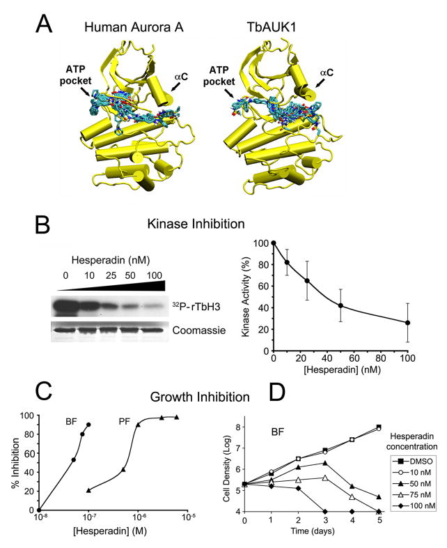
(A) Molecular models of human Aurora A and TbAUK1 were generated based upon the crystal structure of Xenopus Aurora B (2BFY). The top 25 docks to Hesperadin are shown for the models. Each of the bound Hesperadin molecules is represented as a stick figure.
(B) Dose response for Hesperadin inhibition of TbAUK1. The left panel shows an autoradiogram with 32P incorporation into TbH3. The stained Coomassie gel shows that an equivalent amount of TbH3 substrate was loaded onto each gel lane. The right panel shows densitometry of the autoradiograms (n=4; ±SE).
(C) Hesperadin inhibits growth of BF and PF cultures. BF or PF were grown in the presence of increasing concentrations of Hesperadin for 24 hr or 48 hr, respectively. The percent growth inhibition was recorded.
(D) Time course of cell growth in the presence of increasing concentrations of Hesperadin. BF cultures were treated at time 0 with the indicated concentrations of Hesperadin and cell density was followed for 5 days. The limit of detection was 1×10⁴ cells/ml.

