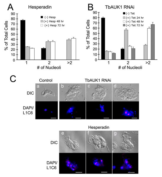
Fig. 7.
Proliferation of nucleoli following treatment with Hesperadin or depletion of TbAUK1 with RNAi.
(A) Nucleoli in Hesperadin treated cells. Cells were treated with 200 nM Hesperadin for the times indicated. The nucleus was stained with DAPI, and nucleoli were labeled with antibody L1C6 and secondary antibodies coupled to Cy3. At least 200 cells were analyzed at each time (n=2; ±SE).
(B) Nucleoli in cells depleted of TbAUK1 by RNAi. The number of nucleoli in TbAUK1 RNAi cells was evaluated at different times after induction with tetracycline. At least 200 cells were analyzed at each time (n=2; ±SE).
(C) Nucleolar labeling with antibody L1C6. Trypanosomes were left untreated (panel a); were examined after 24 hours of induction with tetracycline (+ Tet) (panels b-d); or examined after 24 hr treatment with 200 nM Hesperadin (panels e-g). With RNAi and Hesperadin, note the disruption in nuclear division leading to swollen, multilobed nuclei and multiple nucleoli. The bars are size markers of 10 μm.

