
Figure 1.
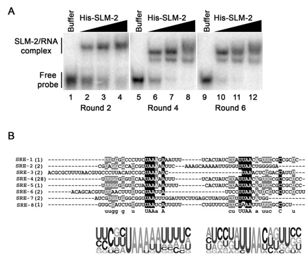
SLM-2 RNA ligands identified. (A) EMSAs of pooled RNAs identified in rounds 2, 4 and 6 using increasing concentrations of His-SLM-2. The protein/RNA complex was separated from the free probe on a native PAGE. The migration patterns of unbound RNAs (free probe) and protein bound RNAs (SLM-2/RNA complex) are indicated on the left. (B) The sequences of 8 unique RNAs bound to SLM-2 after six cycles of SELEX. Both identified motifs are aligned and black undermark. Illustrated, underneath the sequences is the probability matrix (graphic logo) based on all the 8 different sequences, depicting the relative frequency of each residue at each position within the selected motif.