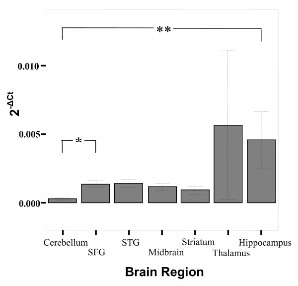
Figure 5.
Bar graph describing the expression (2-ΔCt) of TPH2 in various brain regions. There are significant differences in expression (denoted by ** [p = 0.001] and * [p = 0.011]) across the brain regions, mainly due to lower expression of genes in the cerebellum (see Table 1). Bars represent the mean and error bars represent s.e.m. The groups representing expression in the thalamus have N = 2.

