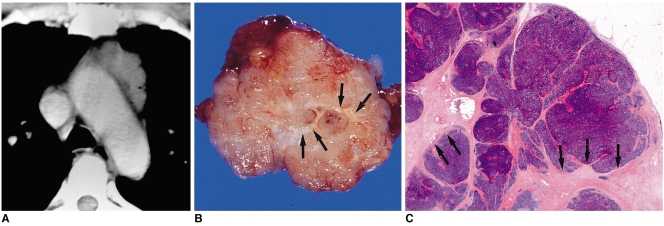
Fig. 4.
Type-B1 thymoma in a 48-year-old-man.
A. Enhanced 10-mm-collimation CT scan obtained at the level of the aortic arch depicts a well-enhanced homogeneous lobulated mass in the left anterior mediastinum.
B. Gross pathologic specimen reveals the presence of well-formed lobules separated by dense fibrous septa (arrows).
C. Photomicrograph (original magnification, × 1; hematoxylin-eosin staining) depicts lobulated internal architecture separated by dense fibrous septa (arrows).

