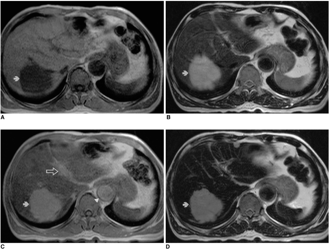
Fig. 1.
Liver hemangioma in a 67-year-old woman.
A. Precontrast T1-weighted in-phase gradient-echo image depicts a round hypointense lesion (arrow) in the right lobe of the liver.
B. Precontrast T2-weighted turbo spin-echo image shows a hyperintense lesion (arrow) in the liver.
C. Ferumoxides-enhanced T1-weighted in-phase gradient-echo image obtained during the distributional phase demonstrates positive enhancement of the lesion (arrow), similar to that of the intrahepatic portal vein (open arrow). Note positive enhancement of the abdominal aorta (arrowhead).
D. Ferumoxides-enhanced T2-weighted turbo-spin echo image shows reduced intensity of the lesion (arrow) compared to precontrast image (A).

