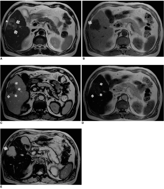
Fig. 3.
Surgically-proven cholangiocarcinoma in segment 5 of the liver.
A. Precontrast T2-weighted turbo spin-echo image shows a heterogeneously hyperintense lesion (arrows) with mild capsular retraction in the right lobe of the liver (arrowhead).
B. Precontrast T1-weighted in-phase gradient-echo image depicts a hypointense mass (arrow).
C. On this T1-weighted out-of-phase gradient-echo image obtained after the administration of ferumoxides, the lesion shows peripheral rim enhancement (arrows).
D. T1-weighted in-phase gradient-echo image obtained after the administration of ferumoxides shows that the lesion (arrows) has become slightly hyperintense to the liver.
E. On this T2*-weighted gradient-echo image obtained after the administration of ferumoxides, the lesion (arrow) has become very hyperintense to the liver. Note that this accumulation phase image provides excellent contrast and lesion conspicuity.

