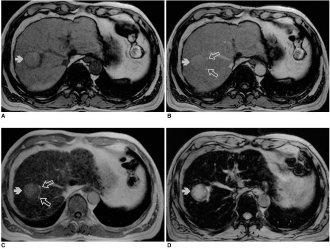
Fig. 4.
A 65-year-old man with biopsy-proven hepatocellular carcinoma in the right lobe of the liver.
A. Precontrast T1-weighted out-of-phase gradient-echo image depicts a hyperintense tumor (arrow) with a hypointense capsule in the right lobe of the liver.
B. Postcontrast T1-weighted out-of-phase gradient-echo image obtained during the distributional phase shows that the lesion (arrow) is hyperintense compared to the liver, but much less hyperintense than the branches of the intrahepatic portal vein (open arrows).
C. Postcontrast T1 in-phase gradient-echo image obtained during the distributional phase shows that the lesion (arrow) has become more hyperintense than the liver but is still less hyperintense than the branches of the portal vein (open arrows). Note the presence of multiple hypointense regenerating nodules in the liver parenchyma.
D. Postcontrast T2*-weighted gradient echo image shows markedly improved lesion (arrow)-to-liver contrast due to the substantially decreased liver parenchymal signal.

