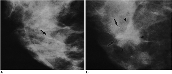
Fig. 1.
A 46-year-old woman with calcifications detected at screening mammography.
A. Routine mammogram obtained at the time of SCNB depicts multiple amorphous round calcifications in a 7-mm cluster, but no mass (arrow). Ultrasonography also failed to identify a mass associated with these calcifications (not shown here). Mammography of the SCNB core tissue specimen revealed a calcified particle, and the histologic diagnosis was fibrocystic change, with calcifications.
B. Mammogram obtained 13 months after SCNB, at which time the patient reported the presence of a lump, reveals a 2-cm-sized, irregular-shaped mass (arrowheads) at the same site, where a similar number of calcifications were present (arrows). Ultrasonography visualized two 2-cm sized masses above and below the nipple (not shown here). A modified radical mastectomy revealed the presence of a 4-cm-sized invasive ductal carcinoma, and single axillary lymph node metastasis.

