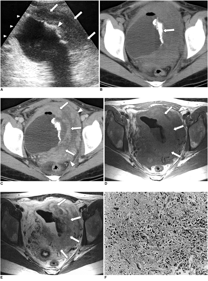
Fig. 1.
A 30-year-old woman with primary lymphoma of the urinary bladder.
A. Ultrasonogram shows diffuse left lateral wall thickening (arrows) and thick linear echogenic foci with posterior acoustic shadowing (arrowhead), suggesting calcification in the left lateral wall.
B. Pre-contrast CT depicts irregular tubular-shaped calcification (arrow).
C. Post-contrast CT demonstrates asymmetric wall thickening (arrows) with heterogeneous enhancement of the left lateral wall. Adjacent to this, displaced small bowel loops (arrowheads) are visible.
D. T1-weighted spin-echo image (TR/TE = 500/8 msec) shows asymmetric thickening of the lateral wall, with low signal intensity (arrows).
E. At T2-weighted fast spin-echo imaging (TR/TE = 3,500/78 msec), high signal intensity is observed (arrows). MR imaging, however, does not reveal the calcification depicted at CT.
F. Photograph (original magnification, ×200; hemotoxylin-eosin staining) of ultrasonography-guided biopsy specimen depicts infiltration of pleomorphic, hyperchromatic lymphoma cells, together with T-cell phenotype and focal necrosis (N).

