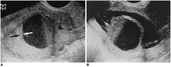
Fig. 5.
A 48 year-old premenopausal woman presented with dysfunctional uterine bleeding.
A. Longitudinal image of transvaginal ultrasonography shows hematometra with a fluid-debris level, and a cervical stenosis is suspected. A thin endometrium (arrows) is seen, which looks normal.
B. Longitudinal image of hysterosonography shows a large endometrial mass, which is near totally necrotic. Total hemorrhagic degeneration of a submucosal myoma was suspected. Hysterectomy was done and adenomyoma was confirmed with pathology testing.

