
FIGURE 4.
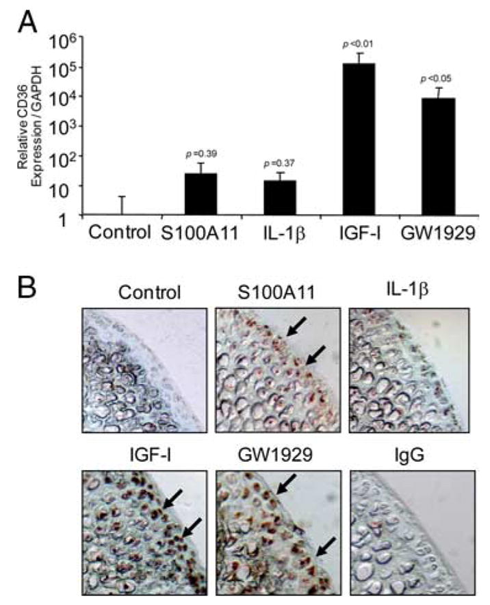
Both IGF-I and the PPARγ agonist GW1929 markedly increase CD36 expression. A, Human immortalized normal knee chondrocytes (CH-8 cells) were stimulated with 100 ng/ml S100A11, 10 ng/ml IL-1β, 10 ng/ml IGF-I, and 100 nM GW1929 for 8 h. Quantitative real-time PCR analysis of CD36 expression (as described in Materials and Methods) is shown pooled from three separate experiments. The p values indicated are compared with control, nonstimulated CH-8 cells. B, Mouse femoral head cartilage explants were stimulated for 48 h with the agonists described in A, and frozen sections were examined by immunohistochemistry for CD36 (arrows) as described in Materials and Methods. Data are representative of five different mouse donors.