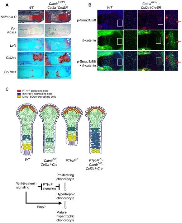
Figure 4. β-catenin may control chondrocyte hypertrophy non-cell autonomously.
Tamoxifen was injected into pregnant females at 13.5 dpc. Embryos were harvested at 17.5 dpc and sections of the proximal tibia were analyzed by Safranin O staining, in situ hybridization with the indicated probes and immnohistochemistry. (A) Upregulated Wnt/β-catenin signaling was indicated by ectopic Lef1 expression and loss of Safranin O staining and Col2a1 expression (arrow). Pictures of higher magnification of the boxed area are shown as insets. Col10a1 expression was not ectopically detected in chondrocytes that had lost Col2a1 expression. (B) Increased p-Smad1/5/8 staining (red) was observed in chondrocyte patches of Catnbex3/+; Col2a1-CreER mouse embryos. Such increase in p-Smad1/5/8 staining was in the area with increased β-catenin staining (green). But p-Smad1/5/8 and β-catenin staining did not colocalize in many chondrocytes. Some cells with increased p-Smad1/5/8 staining (arrows) did not show increased β-catenin staining. (C) Distinct genetic interactions between Wnt/β-catenin and PTHrP signaling in regulating chondrocyte hypertrophy and maturation. The sequential process of chondrocyte hypertrophy and maturation in developing wild type and indicated mutant long bone cartilage were shown in the diagram in the upper panel. Hypertrophic chondrocytes are enlarged and undergo final maturation before they are replaced by the trabecular bone. Initiation of chondrocyte hypertrophy and final maturation are two separate processes that are differentially regulated by Wnt/β-catenin and PTHrP signaling. Wnt/β-catenin signaling controls chondrocyte hypertrophy by inhibiting PTHrP signaling activity. However, Wnt/β-catenin signaling promoted final maturation of hypertrophic chondrocyte independently of PTHrP signaling.

