Fig. 3.
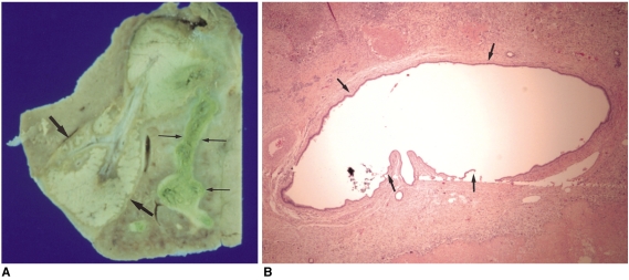
Gross specimen (A) and microphotograph (B) of a rabbit liver resected two weeks (in the late subacute phase) after radiofrequency ablation with the Pringle maneuver.
A. Gross specimen shows tortuous dilatation of the bile duct (thin arrows) adjacent to the ablation zone (arrows).
B. Microphotograph (H & E, ×40) shows the markedly dilated bile duct (arrows).

