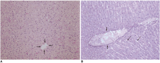
Fig. 5.
Microphotographs (H & E, ×100) of the resected specimens obtained immediately after radiofrequency ablation. The microphotograph of the specimen ablated with the Pringle maneuver (B) shows the distended central vein (arrows) and sinusoids (thin arrows), suggesting congestion, while the central vein (arrows) and sinusoids are normal in the liver ablated without the Pringle maneuver (A).

