Fig. 1.
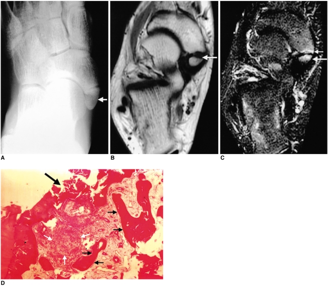
Painful accessory navicular bone in a 14-year-old male soccer enthusiast.
A. Anteroposterior radiograph of right foot shows a type II accessory navicular bone (arrow).
B. Axial T1-weighted spin-echo image (TR/TE, 600/15) shows focal low signal intensity in the medial margin of the accessory navicular bone (arrow).
C. Axial fat-suppressed T2-weighted fast spin-echo image (TR/TE, 4000/132) shows high signal intensity in the accessory navicular bone (long solid arrow), synchondrosis and navicular tuberosity (short solid arrow), which is most intense adjacent to the synchondrosis. At surgery, posterior tibial tendon degeneration was observed.
D. Photomicrograph of excised accessory navicular bone shows destruction of the cartilage cap that represents the synchondrosis (large black arrow), subchondral osteonecrosis (short black arrows) and granulation tissue (white arrows) (H & E stain; original magnification, ×40).

