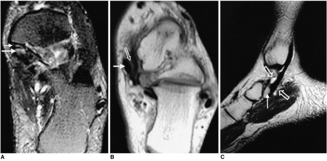
Fig. 2.
Surgically proven synovitis with painful accessory navicular bone in a 16-year-old boy.
A. Axial fat-suppressed T2-weighted fast spin-echo image (TR/TE, 4000/108) shows high signal intensity in the accessory navicular bone (long solid arrow) and synchondrosis (short solid arrow).
B. Axial T1-weighted spin-echo image (TR/TE, 600/11) at a level close to the accessory navicular bone shows a posterior tibial tendon (solid arrow) of normal size and signal intensity. Note the decreased signal intensity around the tendon (open arrow)
C. Sagittal T2-weighted fast spin-echo image (TR/TE, 4000/95) shows fluid in the left posterior tibial tendon sheath (open arrows) with accessory navicular bone (solid arrow).

