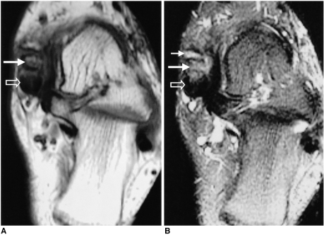
Fig. 3.
Surgically proven partial tear of the posterior tibial tendon with painful accessory navicular bone in a 51-year-old woman.
A. Axial T1-weighted spin-echo image (TR/TE, 500/12) shows low signal intensity in the accessory navicular bone (solid arrow) with distraction of the synchondrosis. The posterior tibial tendon is thickened with increased signal intensity in the tendon (open arrow).
B. Fat-suppressed T2-weighted fast spin-echo image (TR/TE, 4000/108) shows high signal intensity in the accessory navicular bone (long solid arrow) and fluid signal intensity in the synchondrosis (short solid arrow). The posterior tibial tendon displays increased signal intensity (open arrow), indicative of partial thickness tear.

