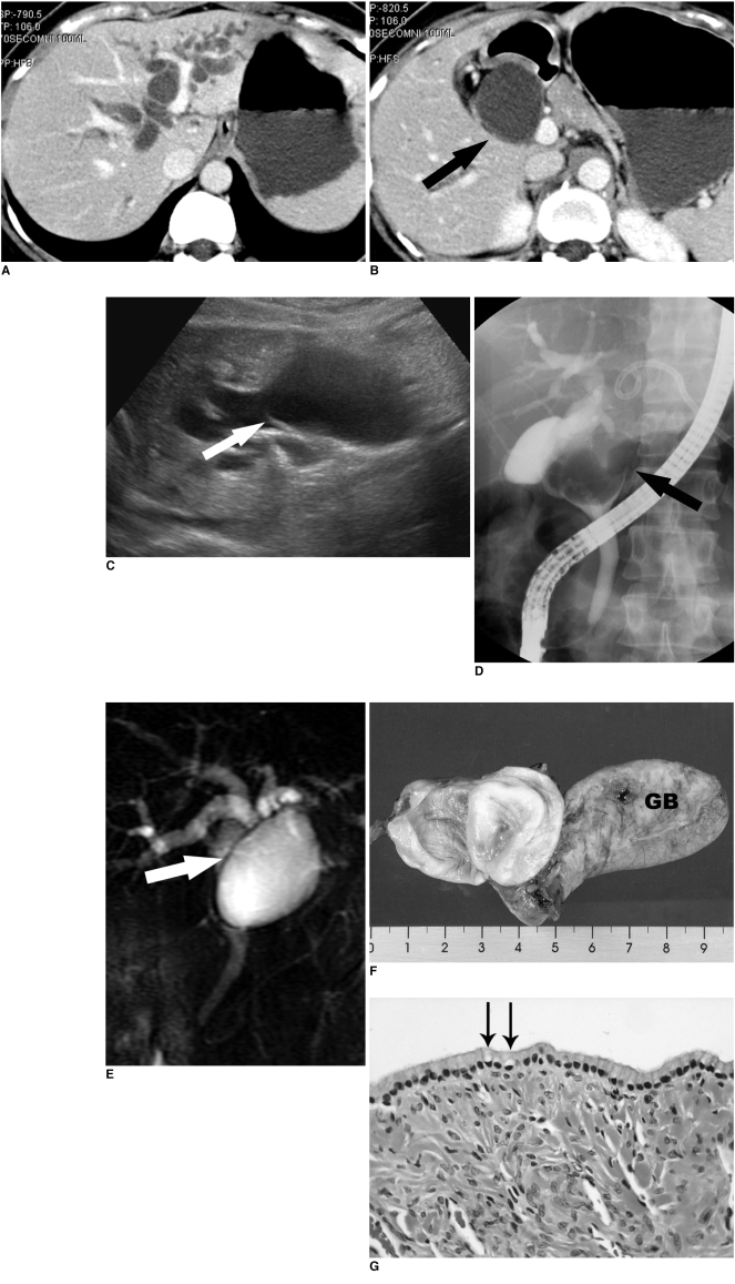
Fig. 1.
Unilocular extrahepatic biliary cystadenoma in a 42-year-old woman.
A, B. Abdominal CT scans show marked dilatation of the intrahepatic duct and the cystic dilatation of the common bile duct (arrow).
C. Ultrasonography also shows cystic dilatation of the extrahepatic duct. On retrospective review, the thin partial septum like structure (arrow) is seen at the upper aspect of the cystic dilatation of the extrahepatic duct.
D. Endoscopic retrograde cholangiopancreatography shows a well-demarcated large filling defect (arrow) in the common hepatic duct with dilatation of the intrahepatic duct. It does not communicate with the bile duct. The PTBD tube, which is not filled with contrast material, is also seen in the left hepatic duct.
E. Magnetic resonance cholangiopancreatography clearly shows the thin wall (arrow) of the cystic lesion in the common hepatic duct. It also shows a bile duct variation; the right posterior segmental duct drained into the left hepatic duct.
F. The surgical specimen shows a unilocular cystic mass arising from the common hepatic duct (incised). GB; gallbladder
G. Microscopically (H & E staining, ×200), the cyst wall is lined by a single layer of columnar mucin-secreting cells (arrows). The epithelium is supported by ovarian-like stroma.

