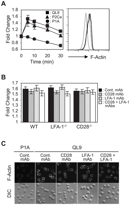
Figure 2. Analysis of F-actin polymerization occurring during culture of 2C T cells with the pMVs.
(A) Wild type (WT) 2C T cells were cultured with the peptide-loaded pMVs for different periods of time and stained for F-actin with FITC-labeled phalloidin followed by flow cytometric analysis. Fold changes in the level of F-actin relative to that of 2C T cells fixed immediately after mixing with the QL9-loaded LdB7-1ICAM-1 pMVs were plotted. Histograms represent the F-actin staining of 2C T cells cultured with P1A- (solid grey), P2Ca- (dotted black) and QL9- (solid black) loaded pMVs for 5 min, respectively. (B) WT, CD28−/− and LFA-1−/− 2C T cells were treated with the respective mAbs, as indicated, before 5 min culture with the QL9-loaded pMVs. Fold changes in the level of F-actin during the culture were plotted. (C) WT 2C T cells were pre-treated with the respective mAbs and cultured with the peptide-loaded pMVs, as indicated, for 20 min on poly-L-lysine-coated coverslips followed by staining for F-actin with FITC-labeled phalloidin.

