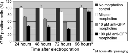
FIG. 1.
Time course of GFP knockdown by morpholinos. Cells were collected at the indicated times after electroporation with water (no-morpholino control), 100 μM mispair anti-GFP morpholino, 10 μM anti-GFP morpholino, or 100 μM anti-GFP morpholino. Fixed samples were subjected to flow cytometry and categorized as GFP positive or GFP negative compared to a wild-type control (see Fig. S1 in the supplemental material). The number of GFP-positive cells in the no-morpholino control culture at each time point was set to 100%. The 96-h culture consisted of cells that were passaged 72 h after electroporation and then grown an additional 24 h before collection. These data are the averages of results for three biological replicates, and error bars represent one standard deviation.

