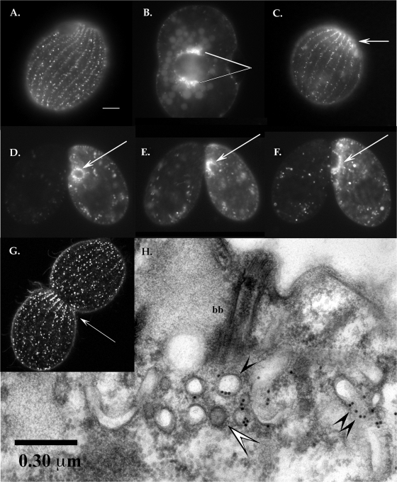
FIG. 3.
Imaging of GFP-tagged Cda12p. Panels A to F are conventional fluorescence micrographs. (A) Live-cell fluorescence image of GFP-Cda12p showing a view of the cell cortex. The signals show up in longitudinal rows of elongated vesicles docked at the cell cortex or mobile “lozenges” traveling both along and between ciliary rows (see the movie in the supplemental material). Scale bar, 10 μm. (B) Deep view showing a dividing cell with GFP-Cda12p-labeled vesicles aggregating at two poles of the macronucleus (pointers). Faint autofluorescence of food vacuoles appears in the background. (C) Postmitotic daughter cell (posterior division product) showing GFP-Cda12p-labeled vesicles concentrated at the fission zone (arrow). (D to F) Mating pairs in which the right partner is labeled with GFP-Cda12p. (D) Fluorescent vesicles are concentrated around the selected postmeiotic micronucleus (arrow). (E) As the selected micronucleus undergoes a third, prezygotic division, the GFP signal labels an anchoring capture cup (arrow). (F) GFP-Cda12p is concentrated around the migratory nucleus (arrow) during pronuclear exchange. (G) Confocal image of a living cell undergoing mitosis. The fluorescent signal is most intense in an area just posterior to the fission zone (arrow), where GFP-Cda12p-decorated vesicles are aggregating. (H) TEM image of GFP-Cda12p-positive vesicles after immunogold labeling. The section through a basal body (bb) shows a rich mixture of membrane-bound vesicles and tubulovesicular compartments. Gold particles appear to decorate noncoated vesicles (single black arrowhead) and tubulovesicular compartments (double black arrowheads). The white arrowhead indicates a coated vesicle.

