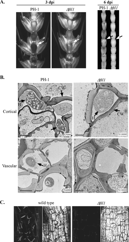
FIG. 3.
The Δftl1 mutant was blocked in colonizing the wheat rachis. (A) Rachises of wheat heads inoculated with the wild type (PH-1) and the Δftl1 mutant strain were examined at 3 (left) or 6 (right) dpi. Wheat kernels on the inoculation side were removed to expose the rachis. The inoculation sites are marked with arrows. (B) TEM images of the cortical (top row) and vascular (bottom row) tissues of the wheat rachis next to the florets inoculated with the wild type (PH-1) or the Δftl1 mutant (Δftl1) at 6 dpi. H, inter- and intracellular hyphae; CW, plant cell wall. Bar, 2 μm. Hyphal growth was only observed in the xylem vessels in wheat heads inoculated with the wild type. (C) Microscopic images of longitudinal sections of rachises and adjacent nodes from wheat heads infected with transformants of the wild type (GZT501) and Δftl1 mutant (RM1) that expressed GFP constitutively. At 6 dpi, intracellular hyphae of the wild type colonized the parenchyma tissue in the rachis. Hyphal growth of the Δftl1 mutant was rare or not observed in the rachis. Left and right panels depict the same areas observed under confocal and differential interference contrast microscopy. A basal level of autofluorescence was visible in the plant cell wall.

