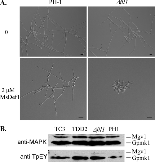
FIG. 5.
Enhanced sensitivity of the Δftl1 mutant to MsDef1. (A) Conidia of the wild-type strain PH-1 (upper panels) and the Δftl1 mutant (lower panels) were incubated in CM for 15 h with 0 or 2 μM defensin MsDef1. Bar, 20 μm. (B) Western blot analyses with proteins isolated from vegetative hyphae of PH-1, Δftl1 mutant T1, LisH domain deletion transformant TDD2, or Δftl1-complemented strain TC1. The upper panel shows detection with the anti-TpEY antibody. The lower panel shows detection with an anti-MAPK antibody.

