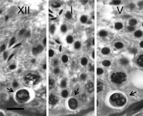
Figure 1.
Photomicrographs of degenerating spermatogonia on the basement membrane of testis from adult rhesus monkeys are shown (arrow) in Stages XII (left), I (middle) and V (right) of the seminiferous epithelial cycle.
Degenerating spermatogonia were typically seen as cells with a small nucleus, characterized by condensed chromatin accumulated at the inner surface of the nuclear membrane surrounding a ground glass-like material. These cells may appear either as solitary (left and middle panel), chains or a coalescence of two or more cells, as shown in the right panel. Bar = 10 µm.

