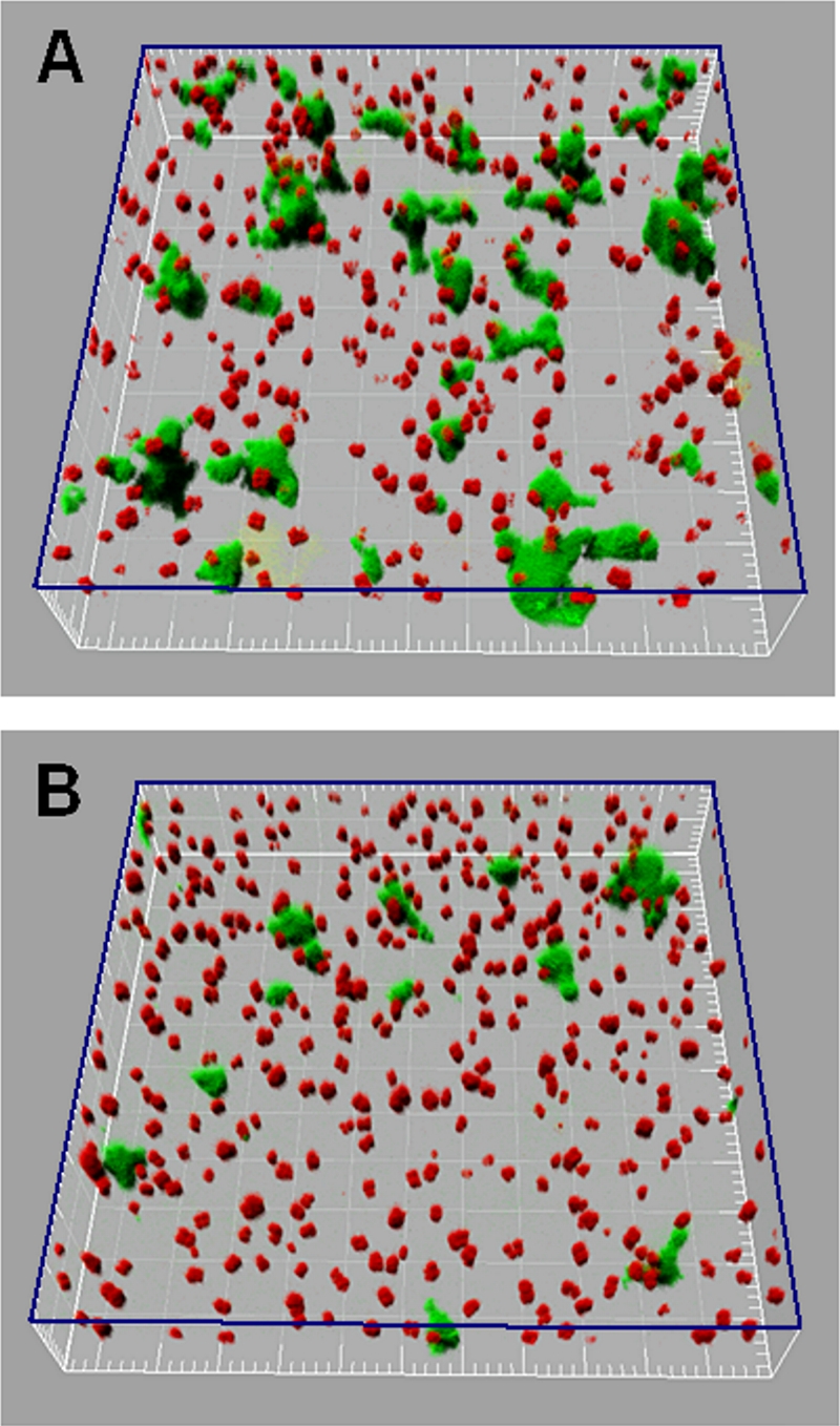
FIG. 6.

Three-dimensional CLSM reconstructions of dual-species biofilms formed after 20 min by continually flowing S. natatoria 2.1gfp cells over glass-surface-attached M. luteus 2.13 cells. (A) Spatial positions of coaggregating S. natatoria 2.1gfp cells that adhered to glass and glass-surface-attached M. luteus 2.13 cells in 10% R2A medium. (B) Spatial positions of the S. natatoria 2.1gfp cells that adhered to glass and often non-glass-surface-attached M. luteus 2.13 cells in 10% R2A medium supplemented with 80 mM galactosamine. The three-dimensional plane indicated in blue represents the glass surface. Green cells are S. natatoria 2.1gfp cells, and red cells are M. luteus 2.13 cells. Dimensions of the boxed regions are 230 μm by 230 μm.
