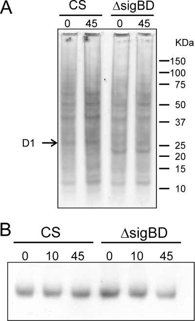
FIG. 7.
Translational activity and amount of the D1 protein in the control and ΔsigBD strains under high light. (A) The cells were pulse-labeled with l-[35S]methionine for 10 min under standard conditions (0 min) and after illumination at a PPFD of 1,500 μmol m−2 s−1 (45 min). Membrane proteins were isolated, separated by sodium dodecyl sulfate-polyacrylamide gel electrophoresis, and blotted onto a membrane for visualization with autoradiography. (B) The amount of the D1 protein was determined by Western blotting with a D1 protein specific antibody.

